HammBurg Be informed with latest news, reviews, entertainment, lifestyle tips, and much more.

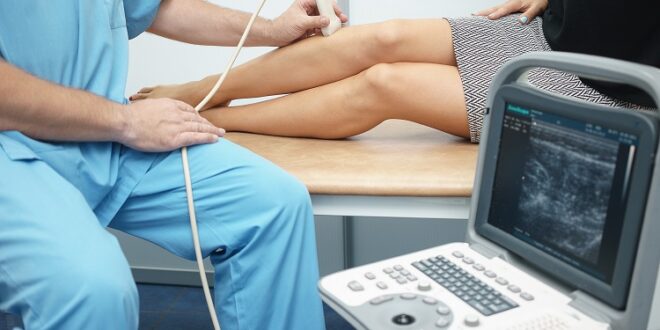
Vascular Ultrasound, which is also referred to as sonography, is the method of producing pictures taken inside our body using soundwaves. A small probe called a transducer produces high-frequency sound waves that travel through the gel that is applied to the body. The probe collects the sound that bounces back from inside the body, and the computer uses these soundwaves to create the image. A vascular ultrasound interpretation course is required to perform this task as these images will be used by physicians to diagnose and treat the ailment.
Uses of Vascular Ultrasound
- It helps in monitoring the blood flow between the organs and the tissues in the body.
- It is used to evaluate varicose veins.
- It helps in preventing aneurysm by determining whether there is an enlarged artery.
- Sonography is used to locate and identify stenosis or blockages, plaques, or emboli so that an accurate venous insufficiency treatment can be identified.
- It detects Deep Venous Thrombosis (DVT) in the body.
- It helps to determine whether a person is accurate for procedures like angioplasty.
- In children, sonography is used to aid the doctors in the placement of a catheter so that major complications like bleeding or nerve injury can be avoided.
- Vascular Ultrasound is also used to identify congenital vascular malformations.
- This procedure is used to identify whether there is an increased blood flow in the patient, which could be a sign of infection.
Procedure
The vascular Ultrasound process is derived from the usage of sonar by bats; that is, when the soundwave hits an object, it bounces back. In the medical world, a transducer is used to send tiny pulses of inaudible and high-frequency soundwaves into the body, and the device records the soundwaves as well. The sound waves bounce off the organs and tissues, and the transducer records the changes in their pitch and direction. These changes are instantly recorded by the computer, which turns the soundwaves into still images or short-loop videos.
Doppler sonography is also performed by the transducer, which measures the speed and direction of the blood cells, and the movement of the cells in the blood vessels causes a change in the soundwaves. This process is called the Doppler effect.
Preparation for the Procedure
Suppose a person is going for vascular ultrasound. In that case, it is advised to wear comfortable and loosely fitted clothes, and they will be asked to remove all clothing and jewelry before entering the examination area. They will be given a gown to wear during the ultrasound. However, in the case of the procedure being conducted on children, they will be provided with distractions throughout the process as they are prone to movements that will prolong the process.
Conclusion
Vascular Ultrasound is a non-invasive process and painless process. While it may be temporarily uncomfortable to the patients, it yields the most fruitful results. It is widely available, cost-effective, and an extremely safe procedure with no harmful effects on humans. A doctor who has undergone a vascular ultrasound interpretation course will be analyzing the images obtained from the procedure. The doctor will suggest follow-up exams to check whether the process has determined the anomaly or not.